|
|||||
 |
 |
 |
|||
 |
|||||
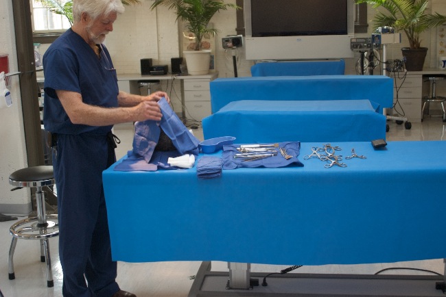
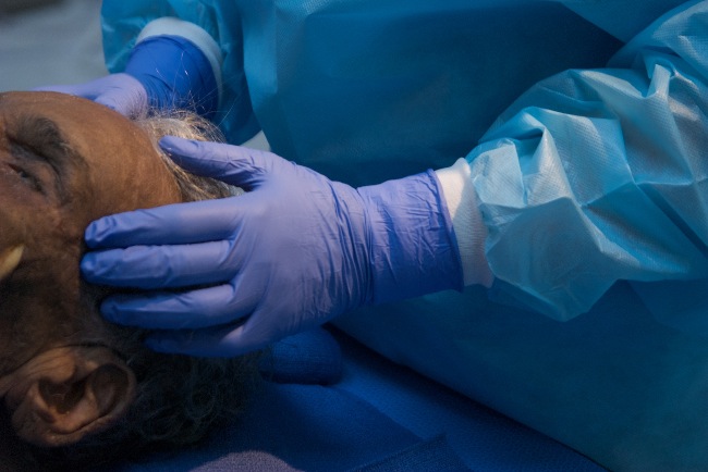
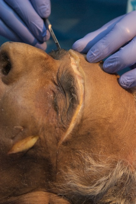
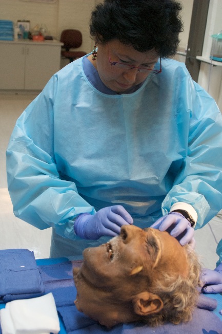
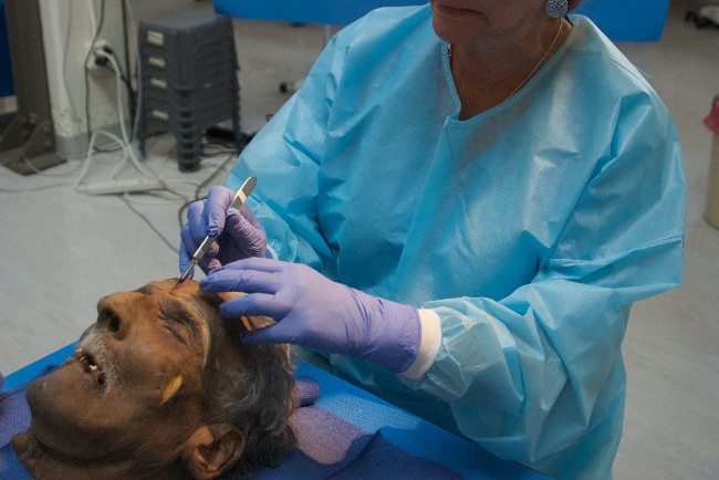
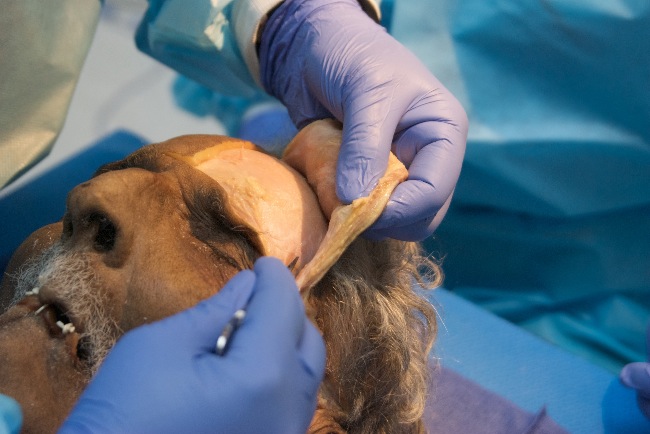
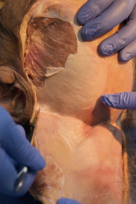
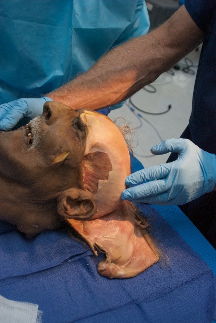
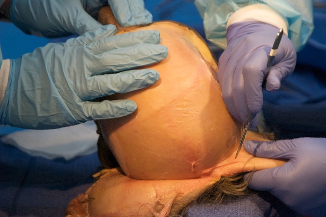
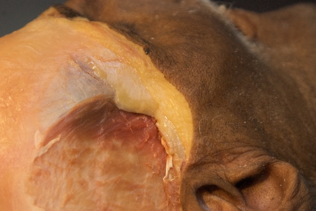
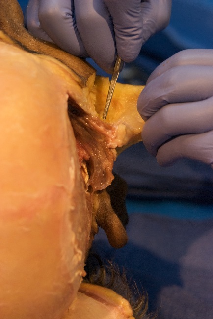
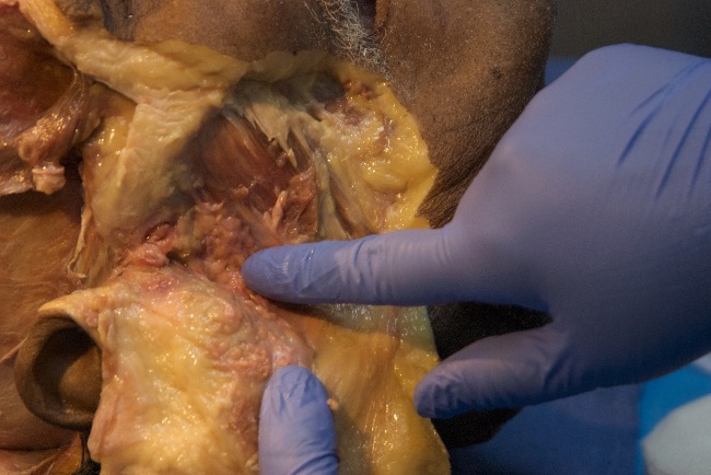
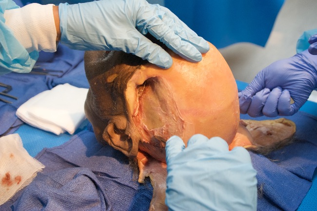
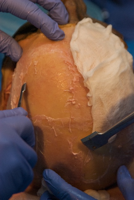
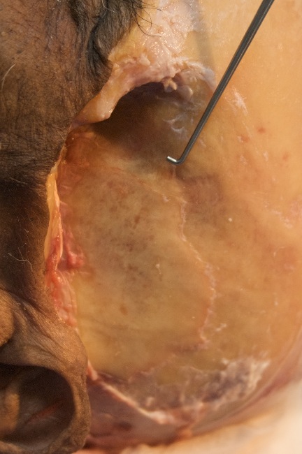
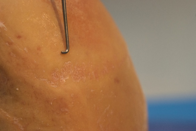
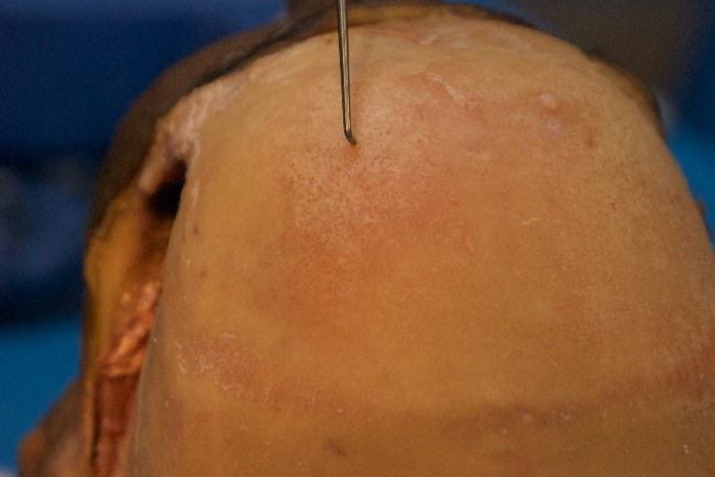
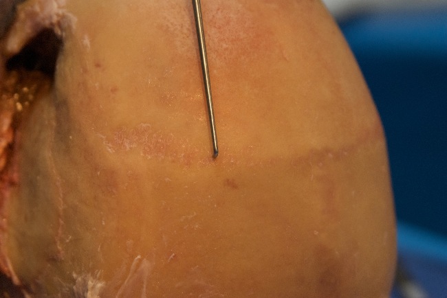
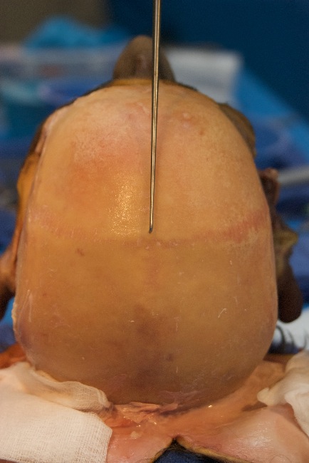
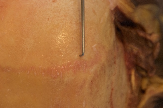
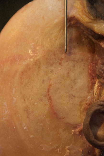
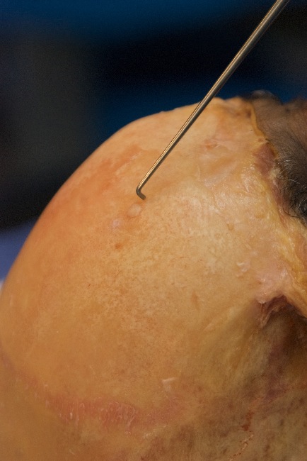
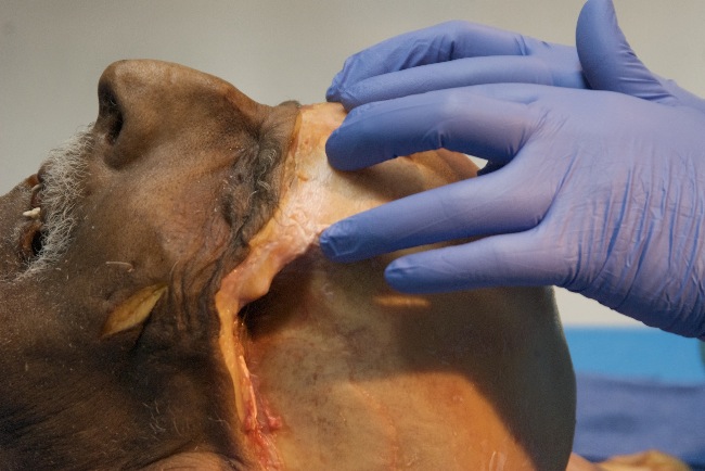
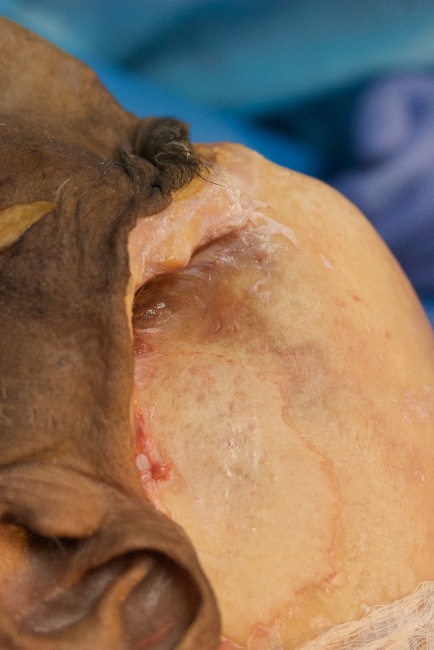
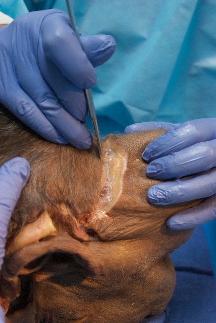
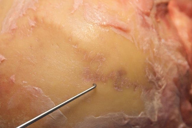
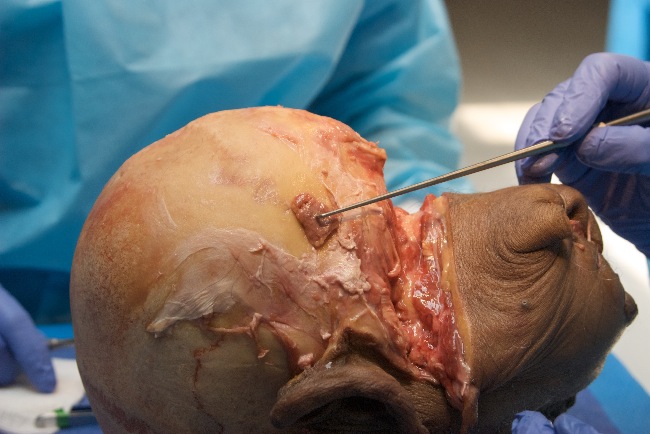
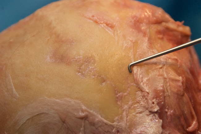
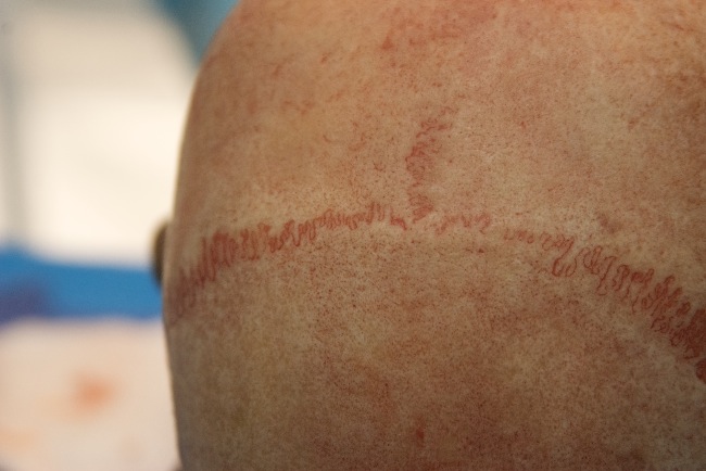
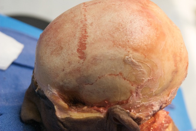
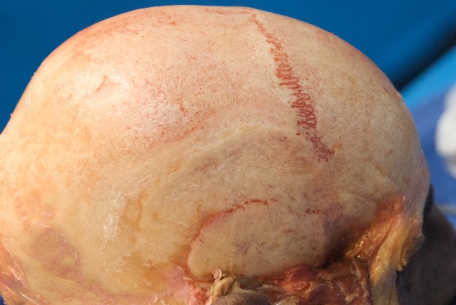
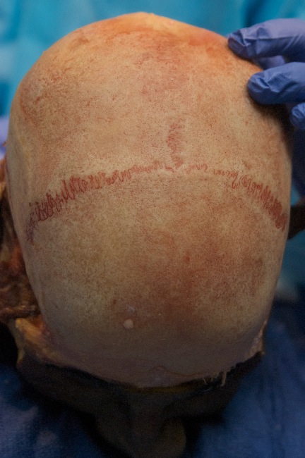
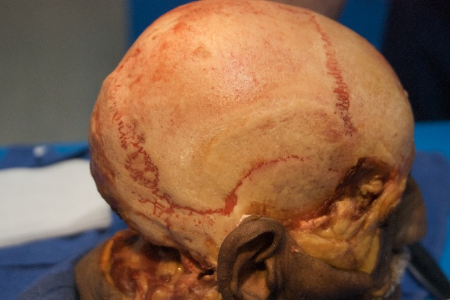
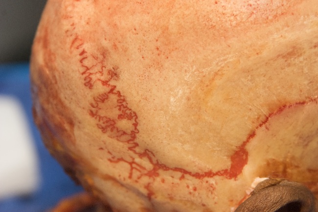
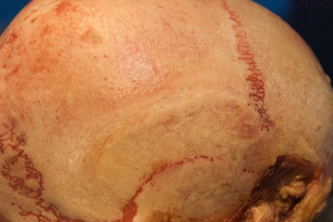
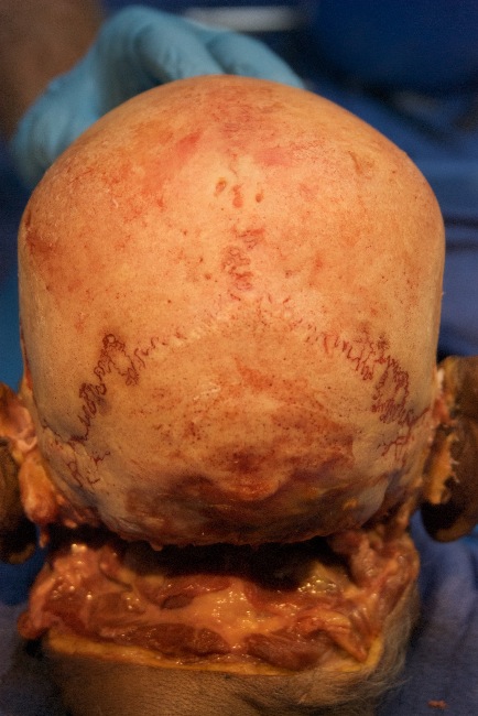
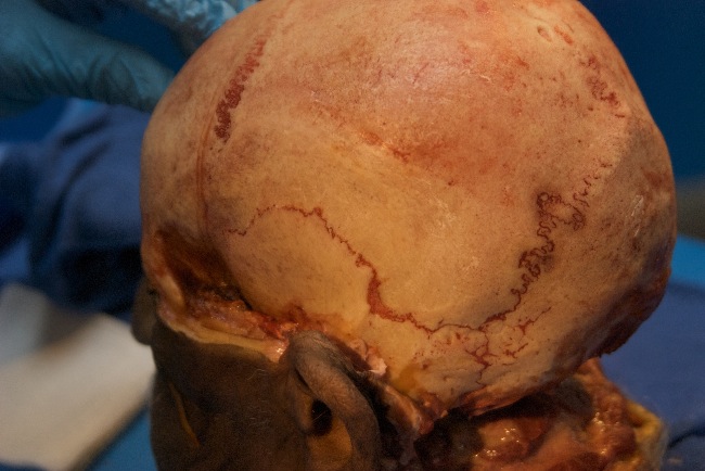
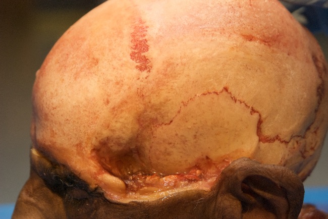
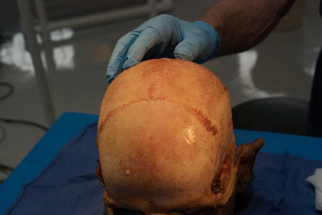
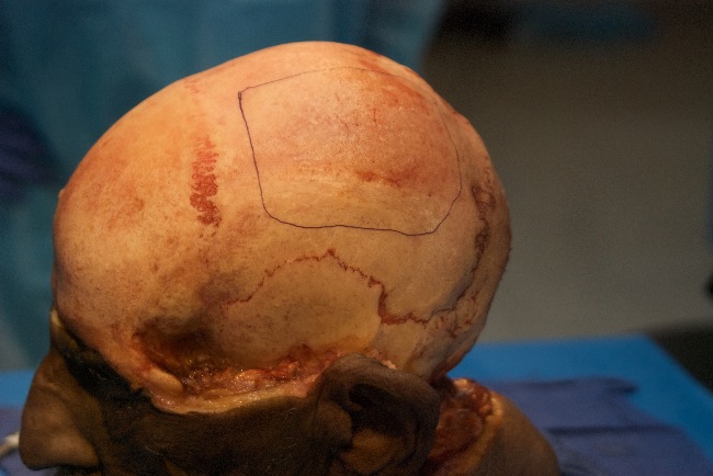
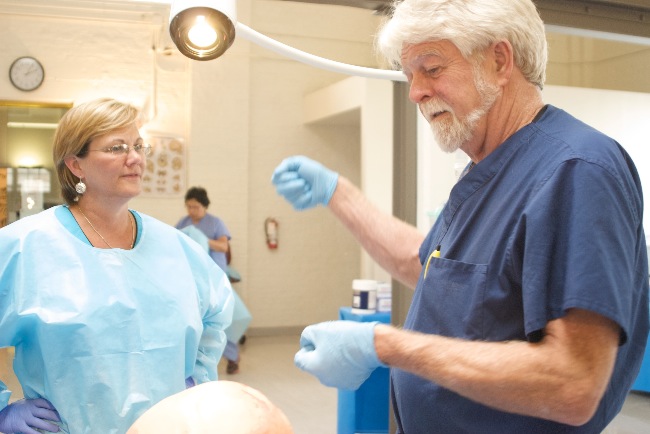
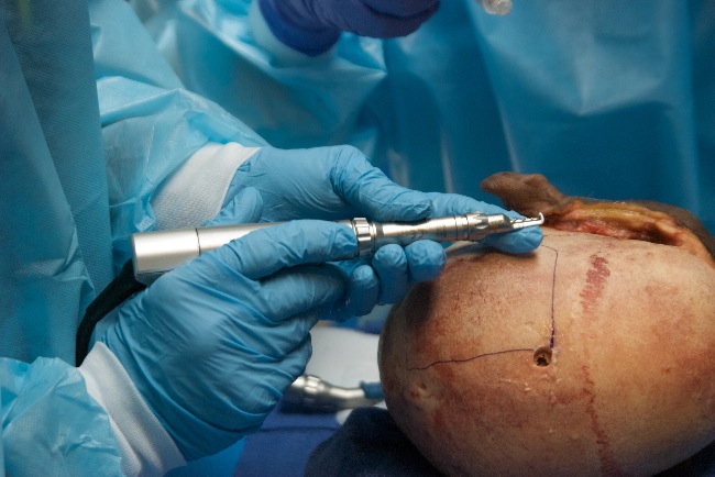
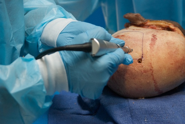
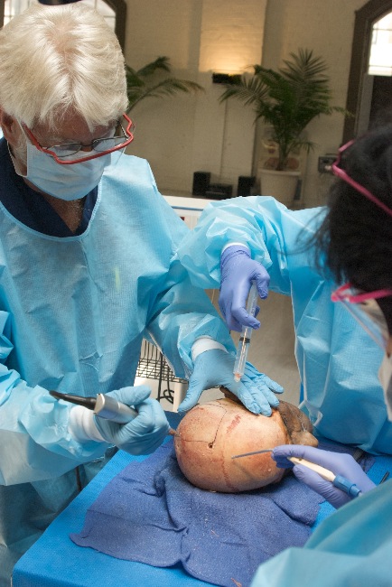
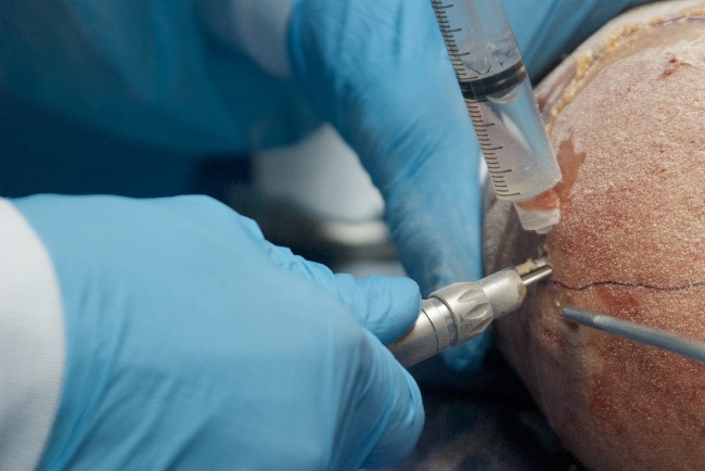
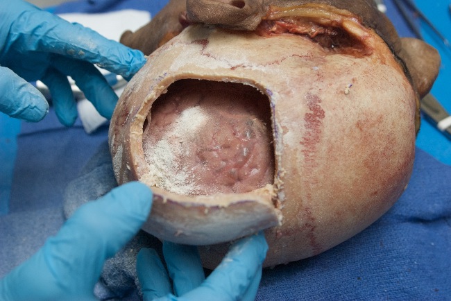
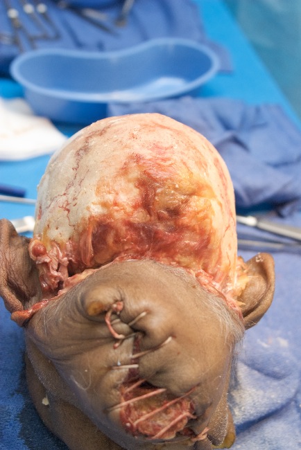
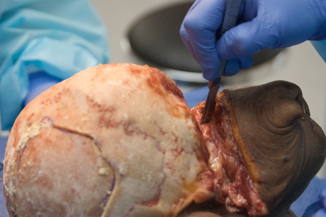
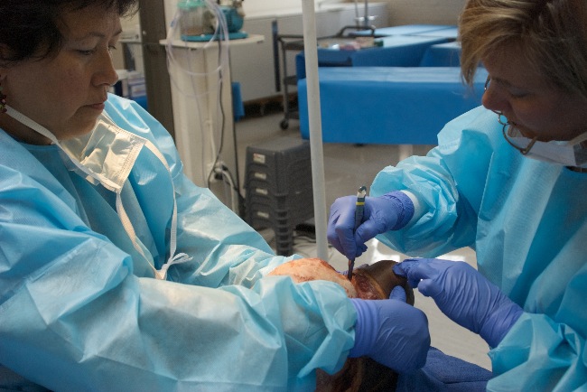
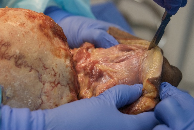
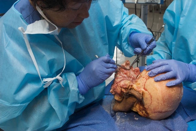
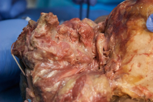
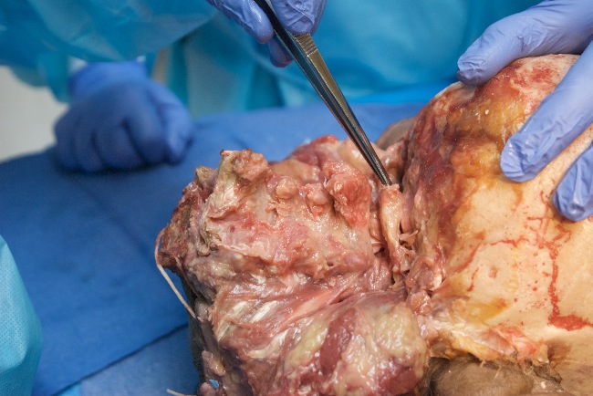
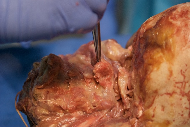
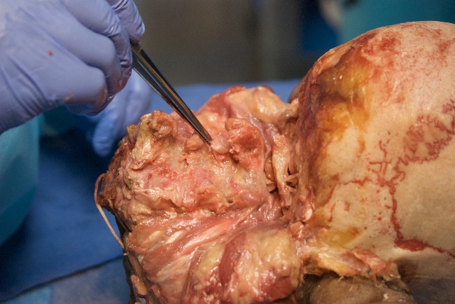
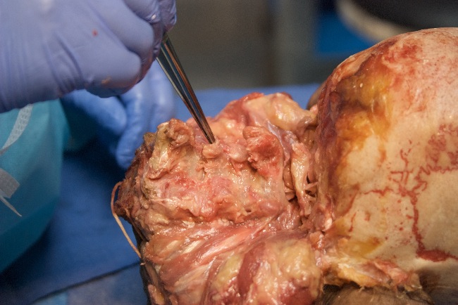
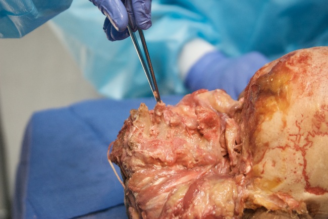
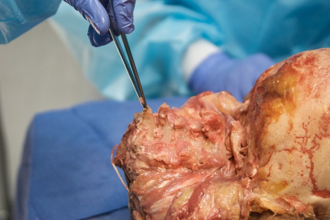
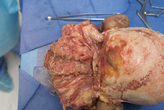
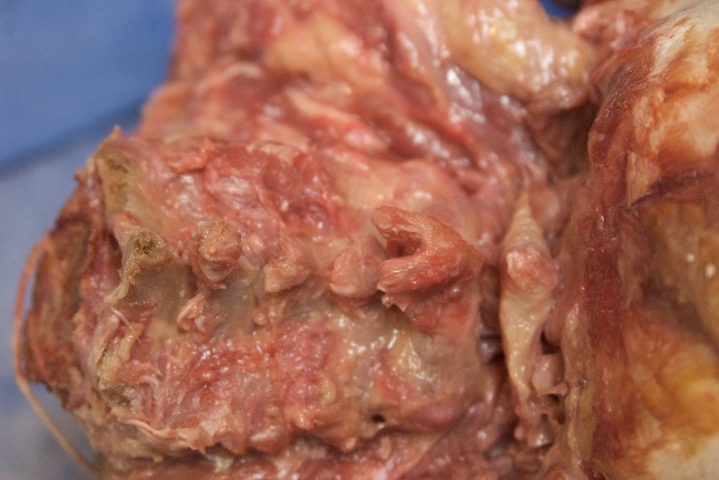
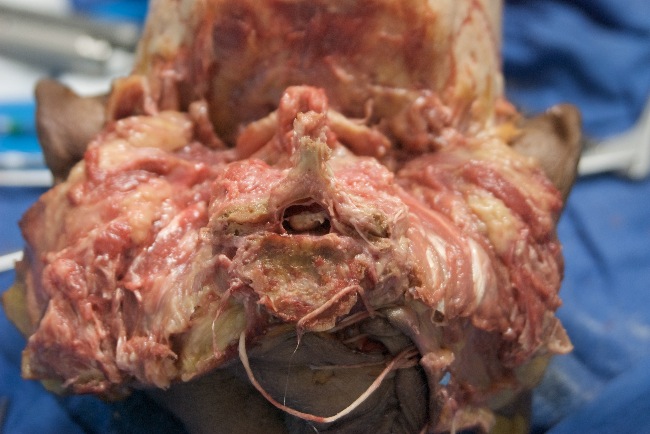
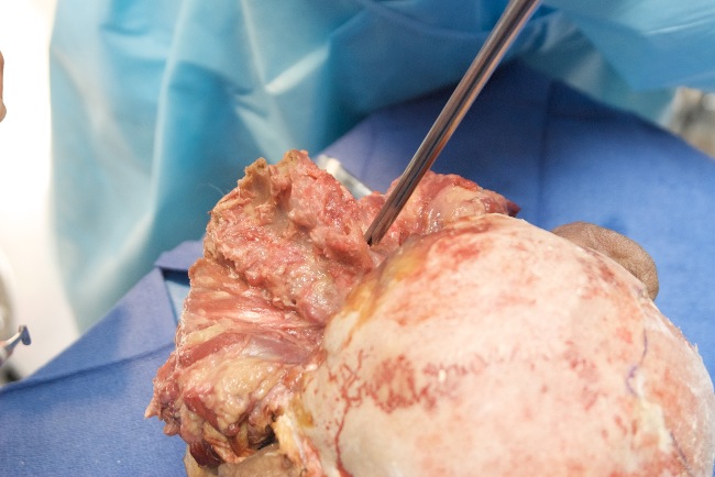
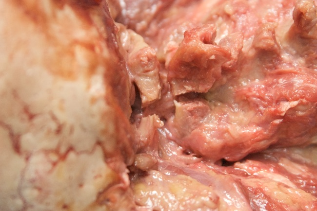
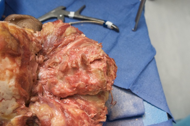
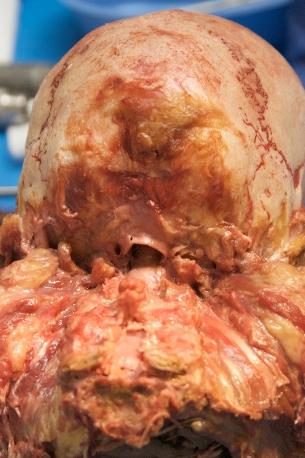
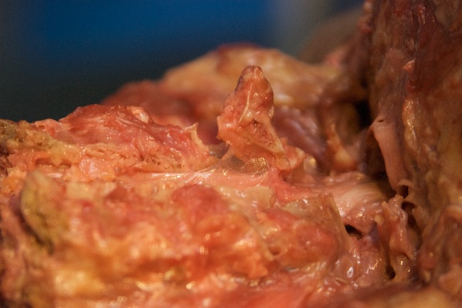
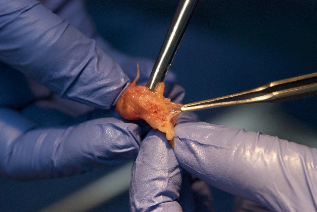
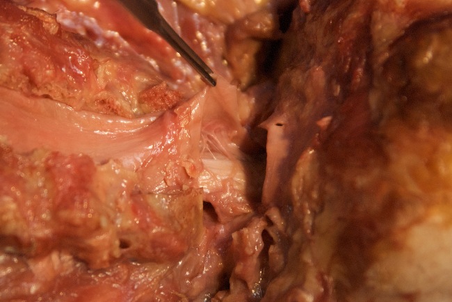
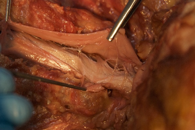
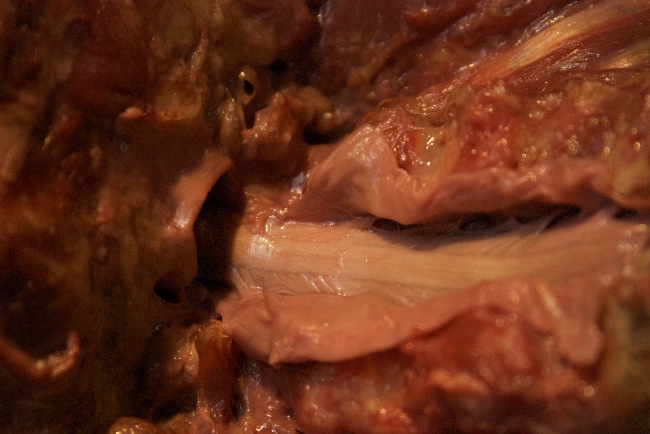
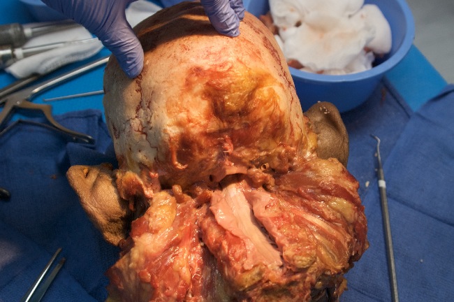
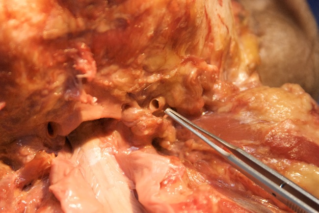
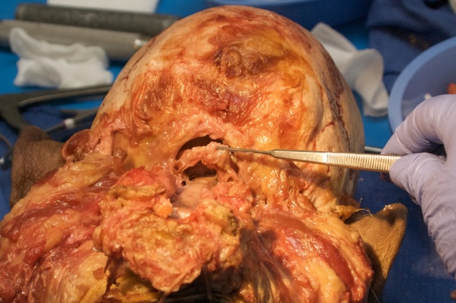
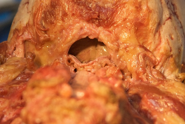
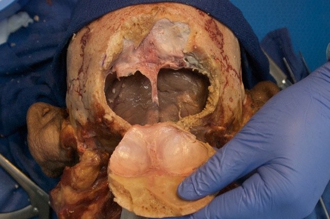
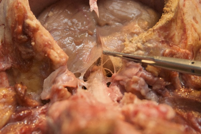
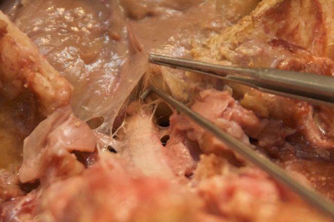
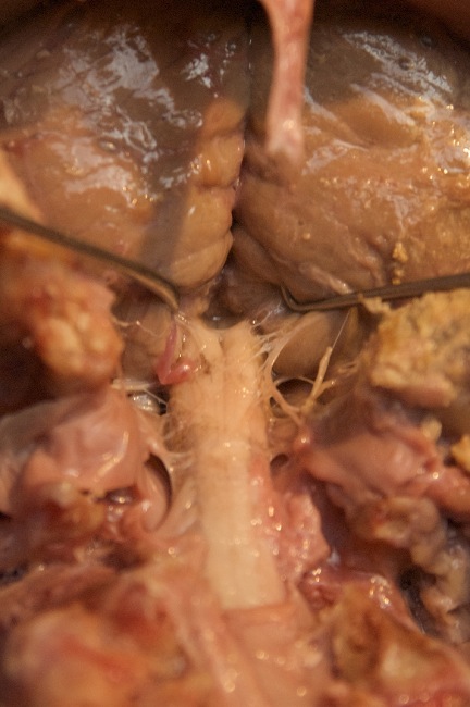
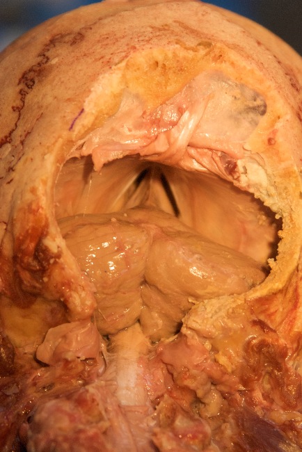
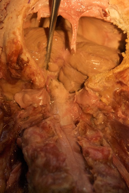
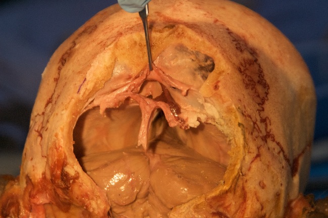
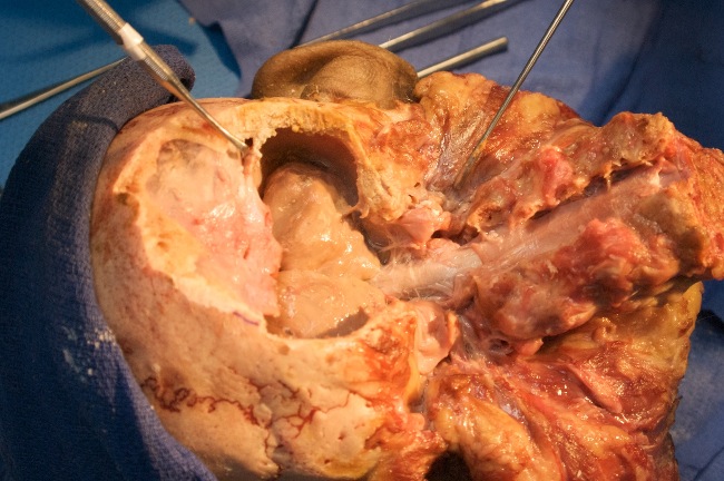
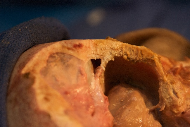
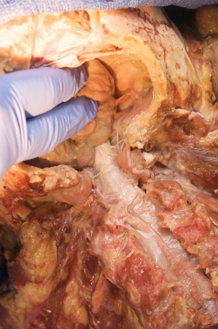
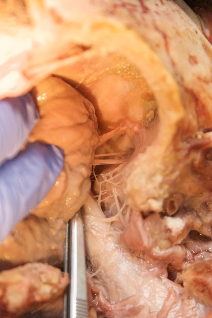
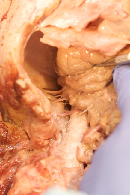
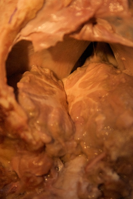
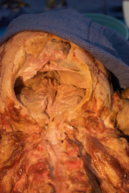
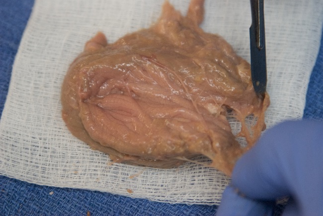
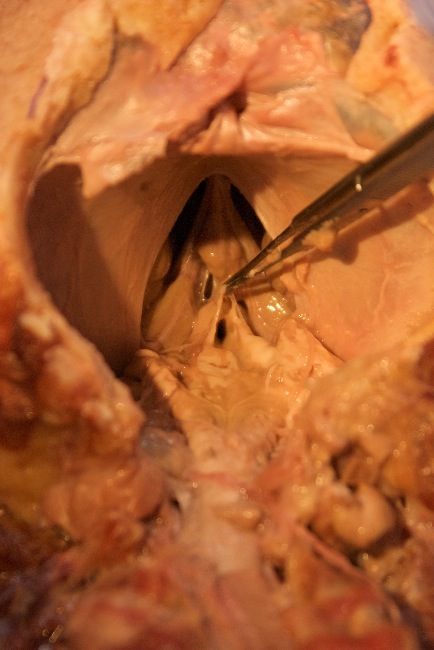
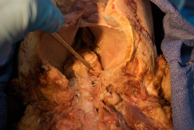
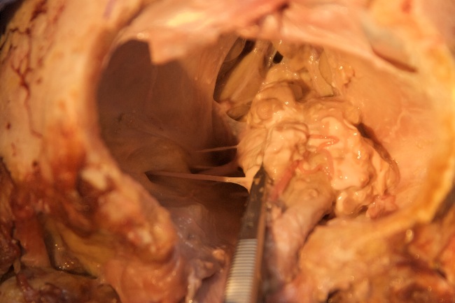
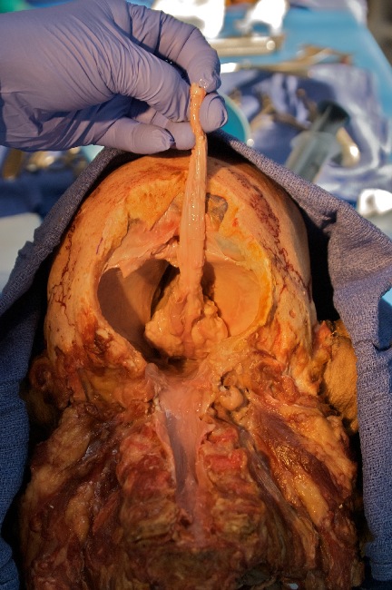
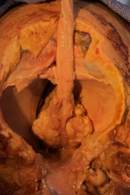
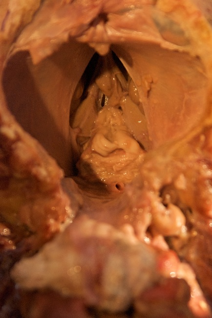
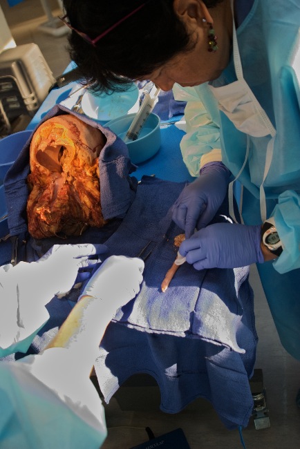
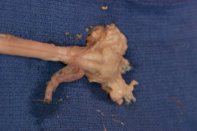
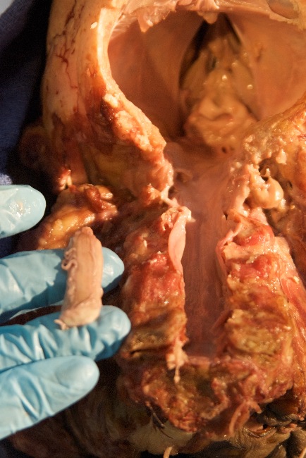
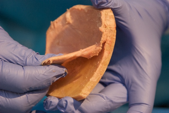
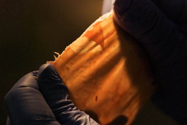
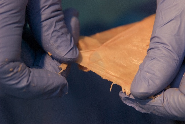
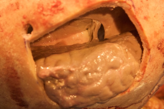
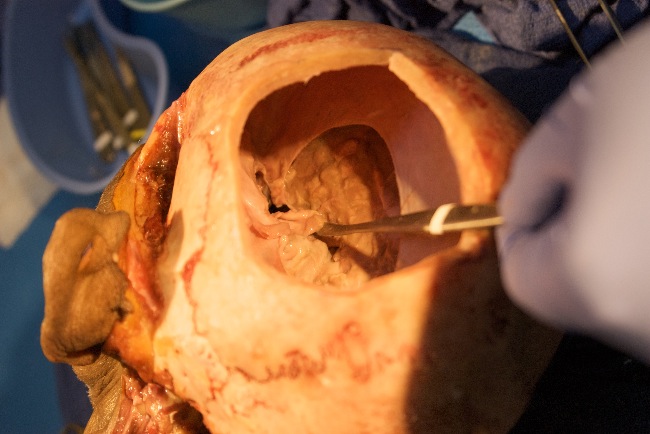
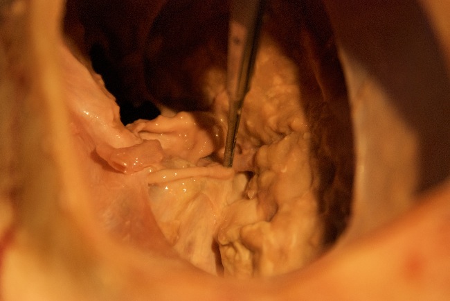
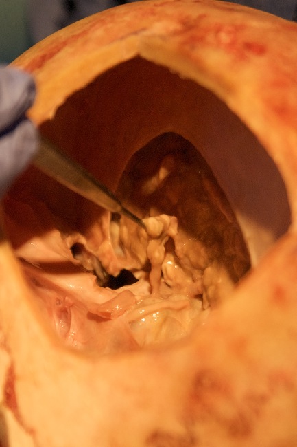
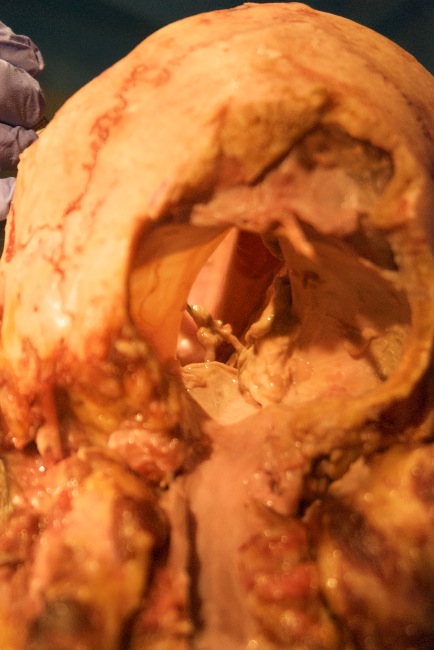
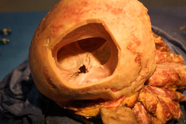
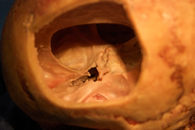
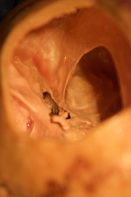
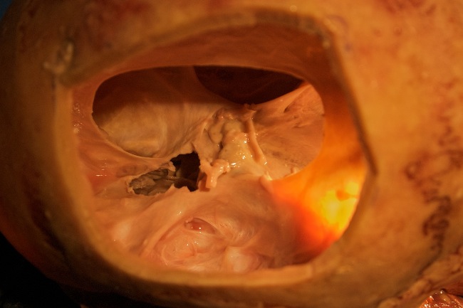
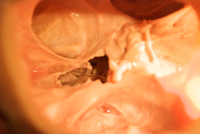
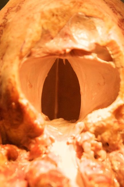
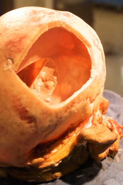
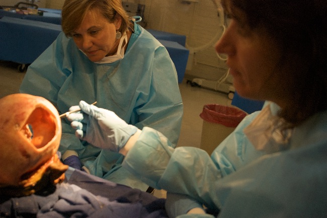
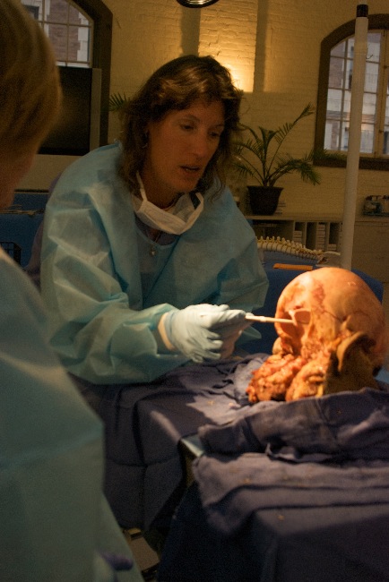
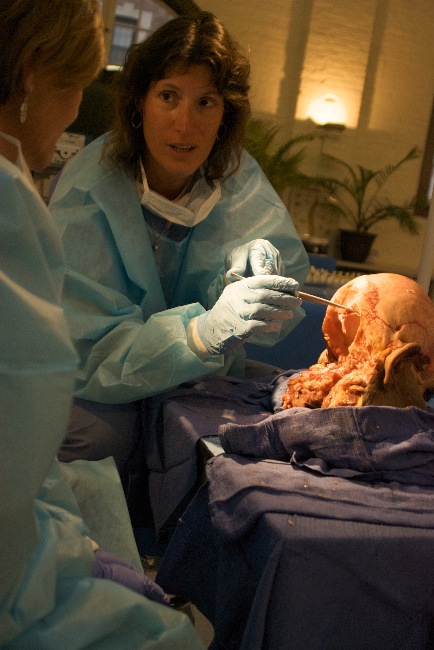
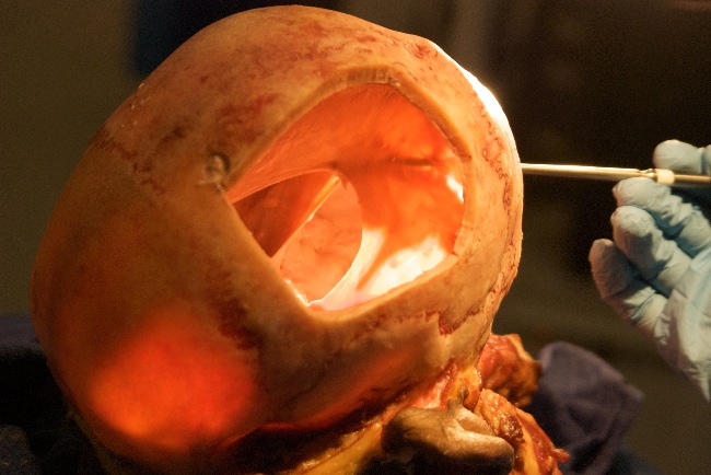
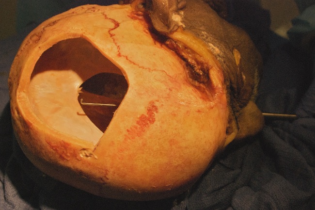
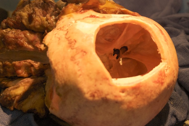
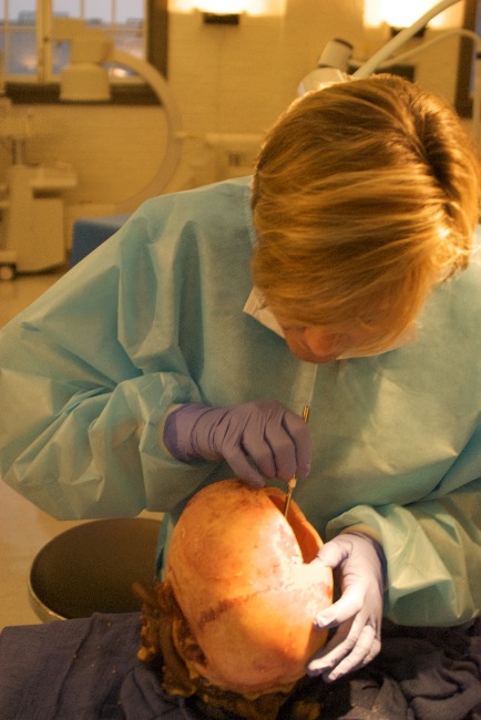
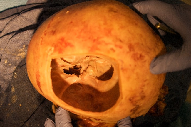
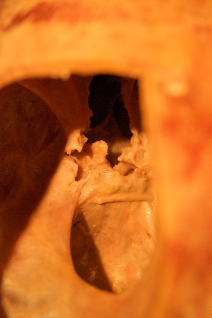
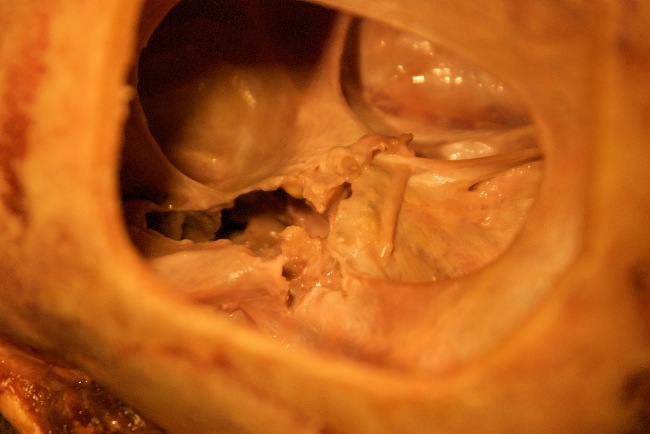
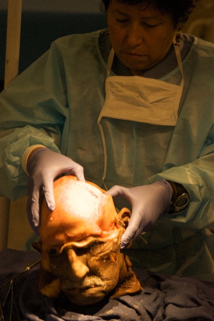
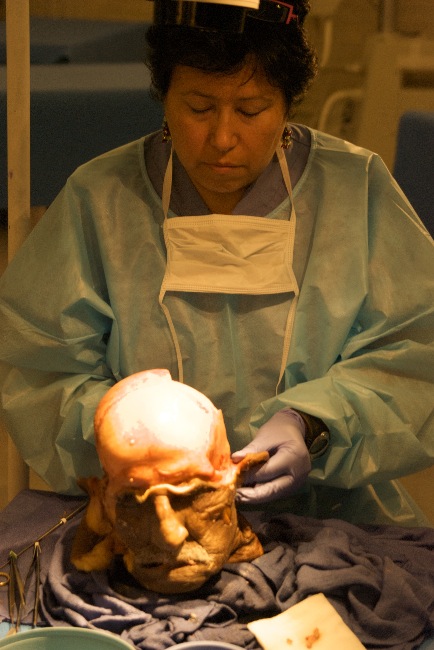
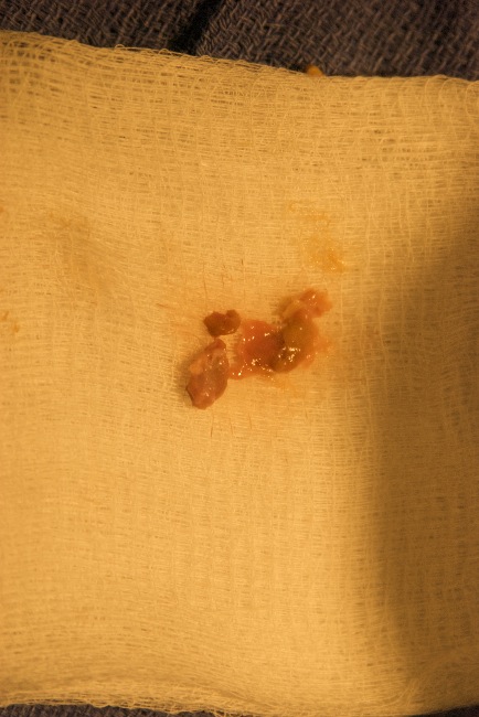
Our specimen during our dissection in August 2010 was one of the most incredible craniums I've had that opportunity to dissect. Shortly after reflecting back the scalp, blood started to collect in the cranial sutures. Yes, I got pictures. You can see in the initial photos the sutures starting to pink up and then they got really bloody red. Bill, the director of VISTA Labs, asked me if I had the cranium in a position that blood was pooling into the sutures. "Nope, the blood came up to the surface. I don't know what the source of the blood was." I informed him. This many not seem amazing but if you consider that most people, including doctors, think that the cranial bones are ossefied at the sutures and don't move, this should give you pause to think about it further. If the cranial bones are ossefied, there would not be any space between the bones for the blood to collect. Because the blood collected in all of the sutures, this is evidence that there is space between the bones and they do not ossefy. Our specimen was in his 90s when he passed away. If his cranial bones hadn't ossified by then, when were they supposed to? In addition to that we found an extra bone growing in his left suboccipital muscles. Deep to where it was located, just superior of the left mastoid process was a wormian bone. (Good thing to look up in Wikipedia or Google if you don't know what it is.) Further investigations revealed a couple of wormian bones along the occipital suture. Our hypothesis is that at a young age, this man, hit the back of his head on the left, which chipped off a small fragment of bone. This bone fragment embedded itself in one of his suboccipital muscles and was nourished with blood and grew. It was about the size of a quarter in diameter and about a quarter inch thick! If that wasn't enough - once we finally opened the cranium we had lots more to explore. The brain tissue was well preserved, especially for a fresh-frozen specimen. We were able to identify many parts of the cortical tissue, brain stem and cranial nerves. Normally we end the day about 4 PM, but we even though it was 6:30 PM were enthralled and wanted to see more! We removed the brain with hands and spoons to examine the intracranial membrane system. As I was explaining its attachments I told the students "The falx cerebri attaches at the sagittal suture, creating the sagittal sinus. It attaches anteriorly at the frontal bone and comes inferior to attach on the crista galli of the ethmoid. You can see the cribiform plates of the ethmoid and vomer here...!" If you knew your cranial anatomy you would know that normally the cribiform plates and vomer are not able to be visualized, unless the ethmoid was cut open. And that was the case here, which explained why we had a difficult time dissecting out the pituitary, as it seemed to be embedded in a lot of scar tissue. Once we removed the pituitary it looked like it was missing a lobe. And we could then see that the anterior aspect of the sphenoid's cella turcica was missing. Putting all these bits of information together we concluded that the man had had a pituitary tumor for which he had surgery. The surgery would have been through the nose or cheek and through the ethmoid and sphenoid. Wow, did he have a lot happen in his life! And because he donated his body to science, we felt so honored to be able to learn about anatomy that we effect with our bodywork techniques, particularly CranioSacral Therapy and NeuroStructural Release. Viewing instructions: Click on an image for a larger view. Put your cursor in the lower right hand corner and click on the "play" arrow to activate the slideshow. Just let me know if you are interested in purchasing a print or an image to use electronically or in print. I'd be happy to talk with you. |
|||

HOME | ABOUT | BOOKS | BLOG | PHOTOS | THERAPY | CLASSES | OTHER | MEDIA | CONTACT | LINKS © 2010 - 2015 Julie McKay Covert Unauthorized use and/or duplication of this material without express and written permission from Julie Covert is strictly prohibited. Excerpts and links may be used, provided that full and clear credit is given to JulieMCovert.com with appropriate and specific direction to the original content. |
||